
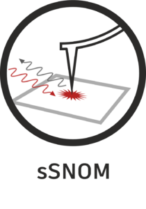
 Scattering-type scanning near-field optical microscopy (s-SNOM or sSNOM) is a powerful, label-free imaging and spectroscopy technique that overcomes the classical optical diffraction limit, achieving nanoscale resolution typically below 50 nm and sometimes down to 10 nm. It combines the sub-nanometer topography capabilities of atomic force microscopy (AFM) with the chemical sensitivity of optical (VIS/IR) spectroscopy. sSNOM utilizes a sharp AFM tip to act as an antenna, focusing light onto a surface and scattering it to enable label-free, sub-diffraction imaging of materials and biological samples. sSNOM employs an “apertureless” approach, whereby a focused laser beam illuminates a metallic or metal-coated AFM tip.
The operating principles can be summarized in a few steps:
Illumination: an external laser beam (visible, IR, or THz) is focused onto the tip apex of a tapping AFM probe. The sharp metallic tip acts as a nano-antenna, concentrating the electric field component of the incident light into a tiny spot (near-field) at the tip apex.
Near-field scattering: the concentrated electric field interacts with the sample surface, inducing scattering that is highly localized to the area directly under the tip. The intensity of the scattering is strongly affected by the local dielectric properties (e.g. refractive index and absorption) of the sample.
Oscillation: because the AFM tip oscillates at a given frequency Ω, the scattering signal is modulated by this oscillation. This allows the detector to separate the highly localized near-field signal from the background far-field signal, which comes from the entire focal spot.
Detection: a lock-in amplifier extracts the signal at the higher harmonics of the oscillation frequency (2Ω, 3Ω,…). This crucial step filters out background noise from far-field reflections, leaving only the localized near-field signal.
Interferometric detection: a modern sSNOM system (such as the one in the SPOTLab) uses an interferometric detection to measure both the amplitude and phase of the scattered light. These relate directly to the local optical properties of the sample, i.e. absorption and reflectivity.
sSNOM is an extremely versatile technique that enables nanoscale infrared (IR) spectroscopy to identify the chemical composition of materials with a spatial resolution of 10 – 20 nm. It is suitable for polymers, organic thin films, and composite materials. It can be used for label-free nano-imaging of living cells, bacteria (e.g. E. coli) and lipids, revealing the distribution of proteins and chemical compositions in aqueous environments. sSNOM is also ideal for mapping surface plasmons in graphene and gold nanostructures, as well as surface phonon polaritons in materials such as hexagonal boron nitride (h-BN). It can identify defects and map free carrier concentrations in semiconductors, as well as monitor phase transitions (e.g. metal-to-insulator transitions) at the nanoscale. Finally, it enables the quantitative determination of the complex optical constants (n, k) of a sample region.
REFERENCES AND APPLICATIONS OF IR NANO-IMAGING AND -SPECTROSCOPY
[1] Biomaterials
[2] Polymers
[3] Inorganic materials
[4] 2D materials