
 Raman spectroscopy is a non-destructive, label-free chemical analysis technique that identifies materials by measuring how they scatter laser light. This scattered light acts as a ‘molecular fingerprint’, carrying information on the Raman-active vibrational modes of the sample and enabling the determination of its chemical structure and composition. Tip-enhanced Raman spectroscopy (TERS) is an advanced, high-sensitivity variant that combines Raman spectroscopy with nanotechnology to map surfaces at the nanometre scale, enabling the visualisation of individual molecules. Both techniques allow for the simultaneous acquisition of images and spectra, yielding hyperspectral data.
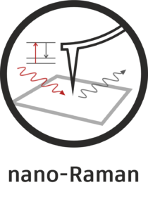
The necessary steps in Raman spectroscopy are:
Laser illumination: a monochromatic laser (typically green or red, e.g. 532 nm or 633 nm for SPOTLab) illuminates the sample.
Inelastic scattering (the Raman effect): most photons scatter elastically (Rayleigh scattering), but a tiny fraction (typically <10-7) scatter inelastically.
Molecular vibrations: inelastic scattering occurs when the light interacts with molecular bonds, more specifically with the vibrational modes of the molecule. This causes the scattered light to gain or lose energy (colour change) equivalent to specific vibrational energy levels.
Spectral analysis: a detector (a spectrograph coupled with a sensitive EMCCD camera) measures this energy difference (the Raman shift), producing a spectrum with peaks that reflect the specific vibrational fingerprint of the molecules.
TERS overcomes the sensitivity and resolution limitations of conventional Raman spectroscopy, which is limited by light diffraction. The above steps apply to TERS, with the following modifications:
A metallic tip antenna is used, which is a very sharp scanning probe microscopy (SPM) tip that is usually coated in gold or silver and positioned within the laser focus.
Plasmonic enhancement: the laser excites surface plasmons (free electron oscillations) at the tip apex, creating an intensely enhanced, localized electric field just a few nanometres wide. This localized field can enhance the Raman signal of molecules immediately beneath the tip by up to 1011 orders of magnitude. The detection stage remains the same as for conventional Raman spectroscopy.
Nanometer resolution: the tip is scanned over the surface, enabling chemical mapping and spectroscopy with a resolution down to 10 – 20 nm or better.
Raman spectroscopy, along with its TERS variant, is useful for identifying unknown substances, contaminants or polymorphs in solids, liquids and gases, and for investigating the uniformity of tablet contents and verifying the quality of raw materials, even when they are contained in blister packs or bottles. It enables chemical mapping and spectroscopy of carbon materials (e.g. graphene and nanotubes), polymers, coatings and semiconductor impurities, and allows for the study of tissue, cells, DNA and proteins without the need for stains or markers (i.e. label-free). TERS takes all these capabilities to another level, enabling single-molecule analysis.
REFERENCES AND APPLICATIONS OF IR NANO-IMAGING AND -SPECTROSCOPY
[1] Polymers
[2] Inorganic materials
[3] 2D materials